Categories
BEAUTY HEALTHY-NUTRITION STRETCHING

Recognizing and preventing sun allergies

photo of a woman with a sunburned face standing in woods and looking skyward, sun is peeking through trees and she is holding her hands at the sides of her face

No one is truly allergic to the sun, but some people are quite sensitive to different types of sun rays and may develop mild to serious reactions after spending time in the sun.

There are several types of “sun allergies,” but polymorphous light eruption (PMLE), an autoimmune condition in the skin that occurs after sun exposure, is one of the most common. Other conditions considered as sun allergies are solar urticaria (hives and reddish patches that usually start 30 minutes to two hours after the sun exposure), actinic prurigo (papules and nodules that are intensely itchy on sun-exposed skin areas), and photoallergic reaction (when the UV rays from the sun modify the chemical structure of medications or products applied to the skin, and a person develops an allergy to the newly modified substance).

What causes PMLE?

People who have PMLE have immune cells that are triggered by sun rays, which attack their skin, and they develop a skin reaction to the sun’s the ultraviolet (UV) rays.

PMLE represents 70% of all sun-induced skin eruptions. It can affect both sexes and all skin types, and it usually starts when someone is a teen or young adult. PMLE may be an inherited condition. Being a female, having fair skin, and living in the north are other risk factors.

PMLE is more common in young women who live in temperate climates. People who live in temperate climates spend all winter out of the sun, so when it becomes warmer the sun exposure is intense. People who live in warmer climates are desensitized because they have a higher sun exposure all year.

What does PMLE look like?

PMLE can appear several hours or days after the first major sunlight exposure of the season, usually during spring or at the beginning of summer. The areas of the body generally affected the most are the ones that are covered during wintertime, but not in the summer: the neck, the chest, and the outer parts of the arms.

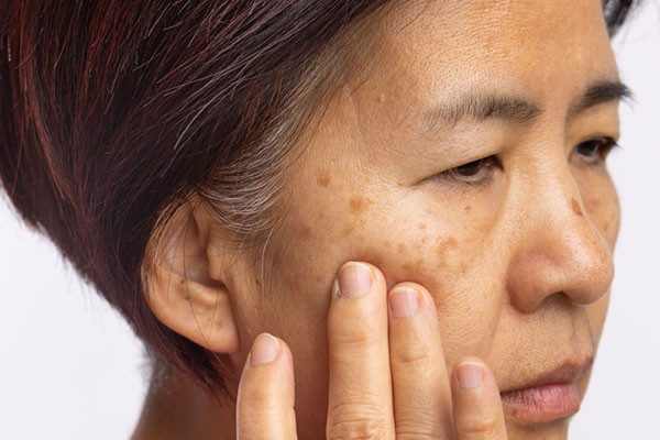
After exposure to the sun, people with PMLE usually notice reddish patches on their skin. These spots may itch, burn, or sting, but they typically don’t leave a scar. In more severe cases, the patches cover most of the body and may also be associated with headaches, fevers, tiredness, and low blood pressure. (If you experience these symptoms, see an urgent care provider for evaluation.) If you think you have PMLE or another sun allergy, a dermatologist is the best doctor to evaluate and treat your skin condition.

Does PMLE get better?

PMLE lesions often get better in approximately 10 days, and it’s important to avoid sun exposure until you are healed. People who develop PMLE can experience significant discomfort and have their life negatively impacted during the spring and summer months. However, repetitive sun exposure can make PMLE less likely to occur. The hardening effect, as it is called, means that the skin lesions that appear after the first episode are less severe, and they can be better tolerated during subsequent episodes.

What are current treatments for any sun allergy, including PMLE?

The best treatment is to prevent sun exposure. Avoid sunlight when it is most intense (from 10 a.m. to 4 p.m.), and use UV-protecting clothing or clothes made of darker and thicker fabrics, as they will prevent the UV rays coming from the sun from reaching your skin. Hats with a wide brim protect your scalp, face, and (partially) the neck.

Broad-spectrum sunscreens that protect your skin from both UVA and UVB rays should be used daily, even if it’s cloudy. Apply sunscreen on your face and any part of your skin that is not covered by a hat or clothing. Reapply sunscreen every two hours, and if you go swimming or get sweaty reapply more frequently (water-resistant sunscreen should also be reapplied).

If you develop PMLE, the areas of skin impacted can be treated with steroid creams. In severe cases, your doctor may recommend a short course of steroid pills. Medications that reduce the immune response, such as azathioprine, are options for treating PMLE, since it is an autoimmune condition (the body is attacking it is own healthy cells).

Antihistamines are medications typically used for allergies that may help shorten the duration of reddish patches that itch or burn, and they also reduce inflammation.

Hydroxychloroquine (a medication also used to treat malaria) can be used in case of flare-ups, or as a prevention method when people travel to sunny locations during winter vacations.

Oral Polypodium leucotomos extract, a natural substance derived from tropical fern leaves, may work as a potent antioxidant, and has anti-inflammatory properties that are beneficial in the prevention of PMLE. Other nutritional supplements containing lycopene and beta-carotene (vitamin A derivatives) have a similar effect. A dermatologist will guide you on the best way to use these medications.

The bottom line

Sun allergies are common in temperate climates, but with a dermatologist’s guidance, vigilant sun prevention, and medications they can be managed throughout the sunny months of the year.

About the Authors

photo of Neera Nathan, MD, MSHS

Neera Nathan, MD, MSHS, Contributor

Dr. Neera Nathan is a dermatologist and researcher at Massachusetts General Hospital and Lahey Hospital and Medical Center. Her clinical and research interests include dermatologic surgery, cosmetic dermatology, and laser medicine. She is part of the … See Full Bio View all posts by Neera Nathan, MD, MSHS photo of Lais Lopes Almeida Gomes

Lais Lopes Almeida Gomes, Contributor

Dr. Lais Lopes Almeida Gomes is a dermatology research fellow at Massachusetts General Hospital, and a pediatric dermatologist in Brazil. Her clinical and research interests include atopic dermatitis and global health. She is part of the … See Full Bio View all posts by Lais Lopes Almeida Gomes

Categories
BEAUTY HEALTHY-NUTRITION STRETCHING

Melasma: What are the best treatments?

close-up photo of a middle-aged woman with spots on her face indicative of melasma, looking concerned and holding her hand to her cheek

Melasma is a pigmentation disorder of the skin mostly affecting women, especially those with darker skin. It is commonly seen on the face, and appears as dark spots and patches with irregular borders. Melasma is not physically harmful, but studies have shown that it can lead to psychological problems and poorer quality of life due to the changes it causes in a person’s appearance.

Melasma is a common disorder, with a prevalence of 1% that can increase to 50% in higher-risk groups, including those with darker skin. Melasma is known as the “mask of pregnancy” since hormonal changes caused by pregnancy, as well as hormonal medications such as birth control pills, are major triggers for excessive skin pigment production in melasma. Sun exposure is another important contributor to melasma.

Can melasma be prevented?

Currently, melasma cannot be fully prevented in people who are likely to develop this condition due to their genetics, skin color type, hormones, or sun exposure level. Avoiding direct sun exposure during peak hours (10 a.m. to 4 p.m.), diligently using high-SPF sunscreens, and avoiding hormonal medications when possible may help protect against melasma flares and reduce their recurrence after treatment. Strict sun protection is the mainstay of any melasma treatment regimen.

What sunscreen should melasma patients use?

Choosing an appropriate sunscreen is critical if you develop melasma, and studies have shown that broad-spectrum tinted sunscreens, especially ones containing iron oxide, can lower pigment production in the skin in melasma patients, as they block visible light as well as UVA/UVB rays. Non-tinted sunscreens, on the other hand, do not block visible light.

For some people, it might be more convenient to use cosmetic products such as foundations that contain both UVA/UVB blockers and visible light blockers such as iron oxide. These products can conceal dark spots and therefore alleviate the psychosocial impact of melasma, and at the same time act as a sunscreen to protect against darkening of the lesions.

It is important for people with melasma to know that visible light can go through windows, and therefore even if they are not out in the sun, they can still get melasma flares by exposing themselves to visible light while driving or sitting by a window.

Can melasma be treated?

Currently there is no cure for melasma; however, there are several medications and procedures available to manage this condition. It is important to know that these treatment options may result in an incomplete response, meaning that some of the discolorations become lighter or disappear while some remain unchanged. In addition, frequent relapses are common.

It is also important to be aware of possible side effects of treatment, including darkening of the skin caused by inflammation induced by the treatment, or extra lightening of the skin in a treated area. Using the appropriate medications under the supervision of a dermatologist can help achieve treatment goals and maintain them with fewer relapses.

Common melasma treatments

The most commonly used treatments for melasma are skin lightening medications that are applied topically. These include medications such as hydroquinone, azelaic acid, kojic acid, niacinamide, cysteamine, rucinol, and tranexamic acid. These medications work by reducing pigment production and inflammation, and by reducing excess blood vessels in the skin that contribute to melasma.

Pregnant women (who constitute a big proportion of melasma patients) should avoid most of these medications except for azelaic acid, which is a safe choice during pregnancy. Hydroquinone is a commonly used skin lightener that should only be used for a limited time due to side effects that may happen with prolonged use. It can be used for up to six months for initial treatment and then occasionally if needed.

In most patients a combination therapy is needed for treatment for melasma. A common choice is the combination of hydroquinone with a retinoid that increases skin cell turnover and a steroid that decreases skin inflammation. Oral medications, including tranexamic acid, are usually considered in more severe melasma cases. This medication is thought to help melasma by reducing pigment production and by reducing excess blood vessels in the skin.

Additional treatment procedures may help

If your melasma does not improve with topical or oral medications, adding procedures such as chemical peels and laser therapies to a treatment regimen could be beneficial.

Chemical peels use substances like glycolic acid, alpha-hydroxy acids, and salicylic acid to remove the superficial layer of the skin that contains excess pigment in melasma patients. The effects of a chemical peel are temporary, since this procedure removes a layer of skin without reducing the production of pigment in regenerating deeper layers.

Laser therapies can destroy pigment cells in skin and therefore lighten the dark spots in melasma. However, as with any other treatment option for melasma, there is considerable risk of relapse post-treatment.

Maintenance therapy and prevention

After achieving improvement of melasma lesions, strict sun protection and maintenance therapy need to be continued. Skin lighteners other than hydroquinone can be used in combination with retinoids to maintain the results, and hydroquinone therapy may be used intermittently if needed.

Takeaway message about melasma

The key point in management of melasma is to use sun protection all the time, and to avoid other triggers such as hormonal medications when possible. Since none of the available treatments are a cure, prevention is the best option. People with melasma should see a board-certified dermatologist for evaluation and appropriate treatment regimens to manage melasma and maintain the treatment results.

About the Authors

photo of Lilit Garibyan, MD, PhD

Lilit Garibyan, MD, PhD, Contributor

Dr. Lilit Garibyan is an assistant professor of dermatology at Harvard Medical School, and a physician-scientist at the Wellman Center for Photomedicine at Massachusetts General Hospital. Her research focuses on innovative biomedical discoveries aimed at identifying … See Full Bio View all posts by Lilit Garibyan, MD, PhD photo of Sara Moradi Tuchayi, MD, MPH

Sara Moradi Tuchayi, MD, MPH, Contributor

Dr. Sara Moradi Tuchayi is a dermatology research fellow at Massachusetts General Hospital. Her research at the Wellman Center for Photomedicine at MGH is focused on the development of novel therapies for skin disorders. See Full Bio View all posts by Sara Moradi Tuchayi, MD, MPH

Categories
BEAUTY HEALTHY-NUTRITION STRETCHING

Should you be tested for inflammation?

A test tube with yellow top is filled with blood and has a blank label. It is lying sideways on top of other test tubes capped in different colors.

Let’s face it: inflammation has a bad reputation. Much of it is well-deserved. After all, long-term inflammation contributes to chronic illnesses and deaths. If you just relied on headlines for health information, you might think that stamping out inflammation would eliminate cardiovascular disease, cancer, dementia, and perhaps aging itself. Unfortunately, that’s not true.

Still, our understanding of how chronic inflammation can impair health has expanded dramatically in recent years. And with this understanding come three common questions: Could I have inflammation without knowing it? How can I find out if I do? Are there tests for inflammation? Indeed, there are.

Testing for inflammation

A number of well-established tests to detect inflammation are commonly used in medical care. But it’s important to note these tests can’t distinguish between acute inflammation, which might develop with a cold, pneumonia, or an injury, and the more damaging chronic inflammation that may accompany diabetes, obesity, or an autoimmune disease, among other conditions. Understanding the difference between acute and chronic inflammation is important.

These are four of the most common tests for inflammation:

  • Erythrocyte sedimentation rate (sed rate or ESR). This test measures how fast red blood cells settle to the bottom of a vertical tube of blood. When inflammation is present the red blood cells fall faster, as higher amounts of proteins in the blood make those cells clump together. While ranges vary by lab, a normal result is typically 20 mm/hr or less, while a value over 100 mm/hr is quite high.
  • C-reactive protein (CRP). This protein made in the liver tends to rise when inflammation is present. A normal value is less than 3 mg/L. A value over 3 mg/L is often used to identify an increased risk of cardiovascular disease, but bodywide inflammation can make CRP rise to 100 mg/L or more.
  • Ferritin. This is a blood protein that reflects the amount of iron stored in the body. It’s most often ordered to evaluate whether an anemic person is iron-deficient, in which case ferritin levels are low. Or, if there is too much iron in the body, ferritin levels may be high. But ferritin levels also rise when inflammation is present. Normal results vary by lab and tend to be a bit higher in men, but a typical normal range is 20 to 200 mcg/L.
  • Fibrinogen. While this protein is most commonly measured to evaluate the status of the blood clotting system, its levels tend to rise when inflammation is present. A normal fibrinogen level is 200 to 400 mg/dL.

Are tests for inflammation useful?

In certain situations, tests to measure inflammation can be quite helpful.

  • Diagnosing an inflammatory condition. One example of this is a rare condition called giant cell arteritis, in which the ESR is nearly always elevated. If symptoms such as new, severe headache and jaw pain suggest that a person may have this disease, an elevated ESR can increase the suspicion that the disease is present, while a normal ESR argues against this diagnosis.
  • Monitoring an inflammatory condition. When someone has rheumatoid arthritis, for example, ESR or CRP (or both tests) help determine how active the disease is and how well treatment is working.

None of these tests is perfect. Sometimes false negative results occur when inflammation actually is present. False positive results may occur when abnormal test results suggest inflammation even when none is present.

Should you be routinely tested for inflammation?

Currently, tests of inflammation are not a part of routine medical care for all adults, and expert guidelines do not recommend them.

CRP testing to assess cardiac risk is encouraged to help decide whether preventive treatment is appropriate for some people (such as those with a risk of a heart attack that is intermediate — that is, neither high nor low). However, evidence suggests that CRP testing adds relatively little to assessment using standard risk factors, such as a history of hypertension, diabetes, smoking, high cholesterol, and positive family history of heart disease.

So far, only one group I know of recommends routine testing for inflammation for all without a specific reason: companies selling inflammation tests directly to consumers.

Inflammation may be silent — so why not test?

It’s true that chronic inflammation may not cause specific symptoms. But looking for evidence of inflammation through a blood test without any sense of why it might be there is much less helpful than having routine healthcare that screens for common causes of silent inflammation, including

  • excess weight
  • diabetes
  • cardiovascular disease (including heart attacks and stroke)
  • hepatitis C and other chronic infections
  • autoimmune disease.

Standard medical evaluation for most of these conditions does not require testing for inflammation. And your medical team can recommend the right treatments if you do have one of these conditions.

The bottom line

Testing for inflammation has its place in medical evaluation and in monitoring certain health conditions, such as rheumatoid arthritis. But it’s not clearly helpful as a routine test for everyone. A better approach is to adopt healthy habits and get routine medical care that can identify and treat the conditions that contribute to harmful inflammation.

About the Author

photo of Robert H. Shmerling, MD

Robert H. Shmerling, MD, Senior Faculty Editor, Harvard Health Publishing

Dr. Robert H. Shmerling is the former clinical chief of the division of rheumatology at Beth Israel Deaconess Medical Center (BIDMC), and is a current member of the corresponding faculty in medicine at Harvard Medical School. … See Full Bio View all posts by Robert H. Shmerling, MD