Categories
BEAUTY HEALTHY-NUTRITION STRETCHING

Untangling grief: Living beyond a great loss

A pink heart on buckling, cracked concrete; concept is broken heart

“The horse has left the barn.”

Those six words, said by my husband’s oncologist, changed our lives forever, although the sense of impending loss had begun weeks earlier with a blood test. There would be more tests, exams, and visits to specialists. As George and I waited for a definitive diagnosis, we bargained with ourselves and with the universe. When we finally met with the cancer treatment team to review all the tests, George’s 6-foot 2-inch frame struggled to fit into the space at the small table, where we strained to follow the conversation. Hearing the word metastatic — meaning cancer had spread throughout his body — was like fingernails on a blackboard.

But there’s no real way to prepare for grief, an inescapable feature of the human condition. Its stress following the death of a loved one can lead to physical illness: cardiovascular diseases, broken-heart-syndrome (takotsubo cardiomyopathy), cancers, and ulcers. Emotional distress often sparks physical distress known as somatic symptoms. How each person navigates grieving varies. Comfort takes different forms for different people. While my journey is individual, my story touches on universal themes, particularly for those grieving in the time of COVID-19.

Anticipatory grief strikes first

George’s diagnosis was advanced metastatic prostate cancer, spread to lymph nodes and bone. There would be no surgery. No radiation. No chemotherapy. Only palliative care.

Some days George wanted to talk only with me. Other days he wanted to talk with those who were “in the same boat.” He saw himself as washed up on the shores of a new, unknown continent. I felt washed up with him. The National Cancer Institute describes these feelings as anticipatory grief, a reaction that anticipates impending loss.

In time, we returned to everyday routines. Sometimes we laughed and didn’t think about his illness. George even conceived of and hosted an annual party for his best friends — men who would be his pallbearers — and their partners. The “pallbearer party,” as it came to be known, was a wonderfully raucous event. Grown men laughed until they cried. Each year, by the end of the night, I knew the tears were for anticipated loss.

George lived another 11 years, more than twice what was expected. But anticipating his loss did not cushion my broken heart.

Acute grief following a death

George died in May 2020, at the beginning of the COVID-19 lockdown. Despite the pallbearers’ dress rehearsals, there was no funeral, no gathering of loved ones. Nothing to soothe my overwhelming pain.

In those first few weeks, time seemed stretched thin, moments repeating themselves like musical notes on a scratched record. I felt untethered, unmoored, adrift. My sides ached from crying; my knees were unsteady. I don’t recall eating.

At the funeral home, when I saw George in a casket, the large room seemed bright from lights hitting the shiny wood floor. Later, I realized the room was much smaller and dimmer than I remembered, its floor not shiny but covered by oriental rugs. Burgundy drapes kept out the sun. As I took in the scene, so different from my recollection, my chest heaved and spasmed.

Such physical reactions and perceptions are common in acute grief. The death of a loved one is accompanied by waves of physical distress that can include muscle aches, shortness of breath, queasy stomach, and trouble sleeping. Food may have no taste, and some experience visual hallucinations. The grief-stricken may not believe their loved one is dead.

Grief in the time of COVID-19

Restrictions to help prevent the spread of COVID-19 disrupted social rituals that connect us during grief. In The Atlantic, Ed Yong describes this absence of much-needed support as the “final pandemic betrayal.”

Although my husband died of cancer, not COVID, I experienced the loss of comforting rituals and the sense that my grief was never truly acknowledged. Experts call this disenfranchised grief. Some predict that prolonged grief disorder driven by this pandemic may reach rates seen only in survivors of natural disasters and wars.

Grief is proof of love

Losing loved ones is not easily incorporated into our life story, though it becomes part of it. The finality and acceptance of a monumental loss takes time. In The Year of Magical Thinking, Joan Didion captures the sudden tragic death of her husband: “John was talking and then he wasn’t.” Life changes in an instant. Yet it takes time to untangle and embrace all that it means.

My life must now be reconfigured and re-envisioned without George. Letting go of grief happens haltingly. Gradually, I noticed that more of my memories of George were happy ones, slowly crowding out the all-consuming early intensity of grief. With time I began to re-engage with the world.

Just as George had, I found I wanted to talk with others in the same boat. A bereavement group helped. I began to exercise more. That helped too. When our dogs died, I got a new puppy. Above all, I learned to be kind to myself.

If you, too, are struggling with loss, experts advise some basics: try to eat, sleep, and exercise regularly; consider a bereavement group or seek out others experiencing grief; stay open to new possibilities — new hobbies, people, and opportunities. Talk to a professional if, after months, you are preoccupied with thoughts of your loved one or find no meaning in life without them. These may be signs that your grief is stalled or prolonged. Effective treatment can help.

Every “first” without George — the first birthday, first wedding anniversary, first anniversary of his death — awakened the early days of intense grief. Still, the experience of living through each made me realize I could survive. I think George would be pleased.

Additional resources

Grief and Loss, CDC

NIH News in Health: Coping with Grief, National Institutes of Health

The Center for Prolonged Grief, Columbia University

About the Author

photo of Martha E. Shenton, PhD

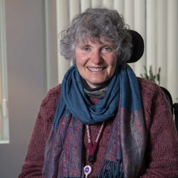
Martha E. Shenton, PhD, Contributor

Dr. Martha Shenton is professor of psychiatry and radiology at Harvard Medical School, and director of the Psychiatry Neuroimaging Laboratory at Brigham and Women’s Hospital in Boston. She and her team have pioneered in developing neuroimaging … See Full Bio View all posts by Martha E. Shenton, PhD

Categories
BEAUTY HEALTHY-NUTRITION STRETCHING

Can a vegan diet treat rheumatoid arthritis?

A brightly colored selection of plant-based vegan foods, including vegetables, fruit, grains, nuts, seeds, and vegan dips.

I recently learned about a study suggesting a vegan diet is an effective treatment for rheumatoid arthritis.

While that sounded intriguing, another claim made in an interview about the study really caught my attention: the lead author of the study said that physicians should encourage people with rheumatoid arthritis to try changing their eating patterns before turning to medication.

Before turning to medication? Now wait just a minute. That flies in the face of decades of research convincingly demonstrating the importance of early medication treatment of rheumatoid arthritis to prevent permanent joint damage. An increasing number of effective treatments can do just that.

In fact, there’s no convincing evidence that changes in diet can prevent joint damage in rheumatoid arthritis. And that includes this new study.

So, what did this research find? Let’s take a look.

A vegan diet for rheumatoid arthritis

Researchers enrolled 44 people with rheumatoid arthritis in the study. All were women, mostly white and highly educated. They were randomly assigned to one of two groups for 16 weeks:

  • Vegan diet. Participants followed a vegan diet for four weeks, followed by additional food restrictions that eliminated foods the researchers considered to be common arthritis trigger foods. These foods included gluten-containing grains (wheat, barley, and rye), white potatoes, sweet potatoes, chocolate, citrus fruits, nuts, onions, tomatoes, apples, bananas, coffee, alcohol, and table sugar. After week seven, these foods were reintroduced, one at a time. Any reintroduced food that seemed to cause pain or other symptoms of rheumatoid arthritis was eliminated for the rest of the 16-week period.
  • Usual diet plus placebo. These participants followed their usual diet and took a placebo capsule each day for 16 weeks. The capsule contained insignificant doses of omega-3 fatty acids and vitamin E.

After the first 16 weeks, participants took four weeks off, then the groups swapped dietary assignments for an additional 16 weeks.

What did the study find about the vegan diet?

The vegan approach seemed to help lessen arthritis symptoms. Study participants reported improvement while on the vegan diet, but no improvement during the placebo phase.

For example, the average number of swollen joints fell from 7 to 3.3 in the vegan diet group, but actually increased (from 4.7 to 5) in the placebo group. In addition, while on the vegan diet, participants lost an average of 14 pounds, while those on the placebo gained nearly 2 pounds.

What else do we need to consider?

While the findings sound great, the study had significant limitations:

  • Size. Only 44 study subjects enrolled and only 32 completed the study. With such small numbers, it only takes a few to alter the results. Larger studies (with several hundred or more participants) tend to be more reliable.
  • Lack of diversity. This trial did not include men and had mostly white, highly educated participants.
  • No standard diagnosis of rheumatoid arthritis. A physician’s diagnosis was required, but there was no requirement that standard criteria be met.
  • Study duration. A treatment lasting four months may seem like a long time, but for a chronic disease like rheumatoid arthritis that can wax and wane on its own, this is too short a time to make firm conclusions.
  • Self-reported diet. We don’t know how well study subjects stuck to their assigned diets.
  • Medication use. Study subjects took arthritis medications, though no information on specific drugs is offered. Some made dosage adjustments during the trial. While the researchers tried to account for this through a separate analysis, the small number of participants could make that analysis unreliable.
  • Weight loss. Losing weight, rather than eating a vegan diet, might have contributed to symptom improvement.
  • No assessment of joint damage. No x-rays, MRI results, or other assessments of joint damage were provided. That’s important, because we know that people with arthritis can feel better even when joint damage continues to worsen. Steroids and ibuprofen are good examples of treatments that reduce symptoms of rheumatoid arthritis without protecting the joints. Without information about joint damage, it’s impossible to assess the true benefit or risk of relying on a vegan diet to treat rheumatoid arthritis.

Finally, it’s unclear how a vegan diet would improve rheumatoid arthritis. This raises the possibility that the findings won’t hold up.

Should everyone with rheumatoid arthritis become vegan?

No, there isn’t enough evidence to justify recommending a vegan diet — or any restrictive diet — for everyone with rheumatoid arthritis.

That said, a plant-rich diet is healthy for nearly everyone. As long your diet is nutritionally balanced and palatable to you, I see little harm in adopting an anti-inflammatory diet. But in the case of rheumatoid arthritis, diet should be combined with medicationto prevent joint damage, not used instead of it.

The bottom line

Growing evidence suggests diet can play a role in treating rheumatoid arthritis. But it’s one thing for a person to feel better on a particular diet; it’s quite another to say diet is enough by itself.

For high cholesterol or high blood pressure, dietary changes are the first choice of treatment. But rheumatoid arthritis is different. Disabling joint damage can occur early in the disease, so it’s important to start taking effective medications as soon as possible to prevent this.

We will undoubtedly see more research exploring the impact of diet on rheumatoid arthritis, other forms of arthritis, and other autoimmune disorders. Perhaps we’ll learn that a vegan diet is highly effective and can take the place of medications in some people. But we aren’t there yet.

About the Author

photo of Robert H. Shmerling, MD

Robert H. Shmerling, MD, Senior Faculty Editor, Harvard Health Publishing

Dr. Robert H. Shmerling is the former clinical chief of the division of rheumatology at Beth Israel Deaconess Medical Center (BIDMC), and is a current member of the corresponding faculty in medicine at Harvard Medical School. … See Full Bio View all posts by Robert H. Shmerling, MD

Categories
BEAUTY HEALTHY-NUTRITION STRETCHING

Ring vaccination might help curtail monkeypox outbreaks

A red figure standing in a white circle connected to a white circle with a black figure, who is connected to circles with two other black figures; the concept of how infection spreads

By now, you’ve probably heard that there is a monkeypox outbreak traveling around the globe. Cases have spread far and wide, including in the US, Canada, Europe, and Australia. It’s the largest outbreak ever recorded outside of western and central Africa, where monkeypox is common.

But controlling this outbreak demands preventive measures, such as avoiding close contact with people who have the illness and vaccination. One method of vaccination, called ring vaccination, has worked well in the past to contain smallpox and Ebola outbreaks. It may be effective for monkeypox as well.

How can monkeypox be contained?

Due to the rising number of cases in multiple countries, the World Health Organization (WHO) has declared monkeypox a global emergency. However, the threat to the public is not considered high. The focus is on identifying possible cases and containing the outbreak as soon as possible.

Three important steps can help stop this outbreak:

  1. Recognize early symptoms
  • Usually, early symptoms are flulike, including fever, fatigue, headache, and enlarged lymph nodes.
  • A rash appears a few days later, changing over a week or two from small flat spots to tiny blisters similar to chickenpox, then to larger, pus-filled blisters.
  • The rash often starts on the face and then appears on the palms, arms, legs, and other parts of the body. If monkeypox is spread by sexual contact, the rash may show up first on or near the genitals.
  1. Take steps to stop the spread
  • Monkeypox spreads through respiratory droplets or by skin-to-skin contact. This includes sexual and other intimate contact.
  •  Anyone who has been diagnosed with monkeypox, or who suspects they might have it, should avoid close contact with others. Once the sores scab over, the infected person is no longer contagious.
  • Health care workers and other caregivers should wear standard infection control gear, including gloves and a mask.
  • In the current outbreak, many cases began with sores in the genital and rectal areas among men who have sex with men, so doctors suspect sexual contact spread the infection. As a result, experts are encouraging abstinence when monkeypox is suspected or confirmed.
  1. Use vaccination to help break the chain
  • Monkeypox is closely related to smallpox. People who received a smallpox vaccine in the past may have some protection from monkeypox. (The US smallpox vaccination program was discontinued in 1972, and smallpox was declared eradicated worldwide in 1980.)
  • Stockpiled smallpox vaccinations and newer vaccines that can be used for monkeypox or smallpox are also available.

Ring vaccination

Monkeypox differs from the virus that causes COVID-19. People with monkeypox usually have symptoms when they’re contagious, and the number of infected persons is usually limited.

This means it’s possible to vaccinate a “ring” of people around them rather than vaccinating an entire population. This selective approach is called ring vaccination.

Ring vaccination has been used successfully to contain smallpox and Ebola outbreaks. It may come in handy for monkeypox as well. Here’s how it works:

  • As soon as a case of monkeypox is suspected or confirmed, the patient and their close contacts are interviewed to identify possible exposures.
  • Vaccination is offered to all close contacts.
  • Vaccination is also offered to those who had close contact with the infected person’s contacts.

Ideally, people should be vaccinated within four days of exposure.

This approach requires widespread awareness of monkeypox, rapid isolation of suspected cases, and an efficient contact tracing system. And of course, vaccines must be available whenever and wherever new cases arise.

Are the vaccines used for monkeypox effective?

According to the CDC, the smallpox vaccine is 85% effective against monkeypox.

While a newer vaccine (JYNNEOS) directed against monkeypox and smallpox has only been tested for effectiveness in animals, it is also expected to be highly effective in humans.

Of course, vaccinations can only work if people are willing to receive them. We’ll learn more about this as more people are offered the option for vaccination.

Are the vaccines used for monkeypox safe?

As with most vaccines, the most common side effects include

  • sore or itchy arm at the site of the injection
  • mild allergic reactions
  • mild fever or fatigue.

Fortunately, more severe side effects, such as significant allergic reactions, are rare.

The bottom line

In light of the current monkeypox outbreak, you may soon be hearing more about ring vaccination. Then again, if appropriate measures are taken to prevent its spread, this outbreak may soon be over. Either way, this won’t be the last time an unusual virus shows up seemingly out of the blue in unexpected places. Climate change, shrinking animal habitats, rising global animal trade, and increasing international travel mean that it’s only a matter of time before this happens again.

About the Author

photo of Robert H. Shmerling, MD

Robert H. Shmerling, MD, Senior Faculty Editor, Harvard Health Publishing

Dr. Robert H. Shmerling is the former clinical chief of the division of rheumatology at Beth Israel Deaconess Medical Center (BIDMC), and is a current member of the corresponding faculty in medicine at Harvard Medical School. … See Full Bio View all posts by Robert H. Shmerling, MD

Categories
BEAUTY HEALTHY-NUTRITION STRETCHING

Recognizing and preventing sun allergies

photo of a woman with a sunburned face standing in woods and looking skyward, sun is peeking through trees and she is holding her hands at the sides of her face

No one is truly allergic to the sun, but some people are quite sensitive to different types of sun rays and may develop mild to serious reactions after spending time in the sun.

There are several types of “sun allergies,” but polymorphous light eruption (PMLE), an autoimmune condition in the skin that occurs after sun exposure, is one of the most common. Other conditions considered as sun allergies are solar urticaria (hives and reddish patches that usually start 30 minutes to two hours after the sun exposure), actinic prurigo (papules and nodules that are intensely itchy on sun-exposed skin areas), and photoallergic reaction (when the UV rays from the sun modify the chemical structure of medications or products applied to the skin, and a person develops an allergy to the newly modified substance).

What causes PMLE?

People who have PMLE have immune cells that are triggered by sun rays, which attack their skin, and they develop a skin reaction to the sun’s the ultraviolet (UV) rays.

PMLE represents 70% of all sun-induced skin eruptions. It can affect both sexes and all skin types, and it usually starts when someone is a teen or young adult. PMLE may be an inherited condition. Being a female, having fair skin, and living in the north are other risk factors.

PMLE is more common in young women who live in temperate climates. People who live in temperate climates spend all winter out of the sun, so when it becomes warmer the sun exposure is intense. People who live in warmer climates are desensitized because they have a higher sun exposure all year.

What does PMLE look like?

PMLE can appear several hours or days after the first major sunlight exposure of the season, usually during spring or at the beginning of summer. The areas of the body generally affected the most are the ones that are covered during wintertime, but not in the summer: the neck, the chest, and the outer parts of the arms.

After exposure to the sun, people with PMLE usually notice reddish patches on their skin. These spots may itch, burn, or sting, but they typically don’t leave a scar. In more severe cases, the patches cover most of the body and may also be associated with headaches, fevers, tiredness, and low blood pressure. (If you experience these symptoms, see an urgent care provider for evaluation.) If you think you have PMLE or another sun allergy, a dermatologist is the best doctor to evaluate and treat your skin condition.

Does PMLE get better?

PMLE lesions often get better in approximately 10 days, and it’s important to avoid sun exposure until you are healed. People who develop PMLE can experience significant discomfort and have their life negatively impacted during the spring and summer months. However, repetitive sun exposure can make PMLE less likely to occur. The hardening effect, as it is called, means that the skin lesions that appear after the first episode are less severe, and they can be better tolerated during subsequent episodes.

What are current treatments for any sun allergy, including PMLE?

The best treatment is to prevent sun exposure. Avoid sunlight when it is most intense (from 10 a.m. to 4 p.m.), and use UV-protecting clothing or clothes made of darker and thicker fabrics, as they will prevent the UV rays coming from the sun from reaching your skin. Hats with a wide brim protect your scalp, face, and (partially) the neck.

Broad-spectrum sunscreens that protect your skin from both UVA and UVB rays should be used daily, even if it’s cloudy. Apply sunscreen on your face and any part of your skin that is not covered by a hat or clothing. Reapply sunscreen every two hours, and if you go swimming or get sweaty reapply more frequently (water-resistant sunscreen should also be reapplied).

If you develop PMLE, the areas of skin impacted can be treated with steroid creams. In severe cases, your doctor may recommend a short course of steroid pills. Medications that reduce the immune response, such as azathioprine, are options for treating PMLE, since it is an autoimmune condition (the body is attacking it is own healthy cells).

Antihistamines are medications typically used for allergies that may help shorten the duration of reddish patches that itch or burn, and they also reduce inflammation.

Hydroxychloroquine (a medication also used to treat malaria) can be used in case of flare-ups, or as a prevention method when people travel to sunny locations during winter vacations.

Oral Polypodium leucotomos extract, a natural substance derived from tropical fern leaves, may work as a potent antioxidant, and has anti-inflammatory properties that are beneficial in the prevention of PMLE. Other nutritional supplements containing lycopene and beta-carotene (vitamin A derivatives) have a similar effect. A dermatologist will guide you on the best way to use these medications.

The bottom line

Sun allergies are common in temperate climates, but with a dermatologist’s guidance, vigilant sun prevention, and medications they can be managed throughout the sunny months of the year.

About the Authors

photo of Neera Nathan, MD, MSHS

Neera Nathan, MD, MSHS, Contributor

Dr. Neera Nathan is a dermatologist and researcher at Massachusetts General Hospital and Lahey Hospital and Medical Center. Her clinical and research interests include dermatologic surgery, cosmetic dermatology, and laser medicine. She is part of the … See Full Bio View all posts by Neera Nathan, MD, MSHS photo of Lais Lopes Almeida Gomes

Lais Lopes Almeida Gomes, Contributor

Dr. Lais Lopes Almeida Gomes is a dermatology research fellow at Massachusetts General Hospital, and a pediatric dermatologist in Brazil. Her clinical and research interests include atopic dermatitis and global health. She is part of the … See Full Bio View all posts by Lais Lopes Almeida Gomes

Categories
BEAUTY HEALTHY-NUTRITION STRETCHING

Melasma: What are the best treatments?

close-up photo of a middle-aged woman with spots on her face indicative of melasma, looking concerned and holding her hand to her cheek

Melasma is a pigmentation disorder of the skin mostly affecting women, especially those with darker skin. It is commonly seen on the face, and appears as dark spots and patches with irregular borders. Melasma is not physically harmful, but studies have shown that it can lead to psychological problems and poorer quality of life due to the changes it causes in a person’s appearance.

Melasma is a common disorder, with a prevalence of 1% that can increase to 50% in higher-risk groups, including those with darker skin. Melasma is known as the “mask of pregnancy” since hormonal changes caused by pregnancy, as well as hormonal medications such as birth control pills, are major triggers for excessive skin pigment production in melasma. Sun exposure is another important contributor to melasma.

Can melasma be prevented?

Currently, melasma cannot be fully prevented in people who are likely to develop this condition due to their genetics, skin color type, hormones, or sun exposure level. Avoiding direct sun exposure during peak hours (10 a.m. to 4 p.m.), diligently using high-SPF sunscreens, and avoiding hormonal medications when possible may help protect against melasma flares and reduce their recurrence after treatment. Strict sun protection is the mainstay of any melasma treatment regimen.

What sunscreen should melasma patients use?

Choosing an appropriate sunscreen is critical if you develop melasma, and studies have shown that broad-spectrum tinted sunscreens, especially ones containing iron oxide, can lower pigment production in the skin in melasma patients, as they block visible light as well as UVA/UVB rays. Non-tinted sunscreens, on the other hand, do not block visible light.

For some people, it might be more convenient to use cosmetic products such as foundations that contain both UVA/UVB blockers and visible light blockers such as iron oxide. These products can conceal dark spots and therefore alleviate the psychosocial impact of melasma, and at the same time act as a sunscreen to protect against darkening of the lesions.

It is important for people with melasma to know that visible light can go through windows, and therefore even if they are not out in the sun, they can still get melasma flares by exposing themselves to visible light while driving or sitting by a window.

Can melasma be treated?

Currently there is no cure for melasma; however, there are several medications and procedures available to manage this condition. It is important to know that these treatment options may result in an incomplete response, meaning that some of the discolorations become lighter or disappear while some remain unchanged. In addition, frequent relapses are common.

It is also important to be aware of possible side effects of treatment, including darkening of the skin caused by inflammation induced by the treatment, or extra lightening of the skin in a treated area. Using the appropriate medications under the supervision of a dermatologist can help achieve treatment goals and maintain them with fewer relapses.

Common melasma treatments

The most commonly used treatments for melasma are skin lightening medications that are applied topically. These include medications such as hydroquinone, azelaic acid, kojic acid, niacinamide, cysteamine, rucinol, and tranexamic acid. These medications work by reducing pigment production and inflammation, and by reducing excess blood vessels in the skin that contribute to melasma.

Pregnant women (who constitute a big proportion of melasma patients) should avoid most of these medications except for azelaic acid, which is a safe choice during pregnancy. Hydroquinone is a commonly used skin lightener that should only be used for a limited time due to side effects that may happen with prolonged use. It can be used for up to six months for initial treatment and then occasionally if needed.

In most patients a combination therapy is needed for treatment for melasma. A common choice is the combination of hydroquinone with a retinoid that increases skin cell turnover and a steroid that decreases skin inflammation. Oral medications, including tranexamic acid, are usually considered in more severe melasma cases. This medication is thought to help melasma by reducing pigment production and by reducing excess blood vessels in the skin.

Additional treatment procedures may help

If your melasma does not improve with topical or oral medications, adding procedures such as chemical peels and laser therapies to a treatment regimen could be beneficial.

Chemical peels use substances like glycolic acid, alpha-hydroxy acids, and salicylic acid to remove the superficial layer of the skin that contains excess pigment in melasma patients. The effects of a chemical peel are temporary, since this procedure removes a layer of skin without reducing the production of pigment in regenerating deeper layers.

Laser therapies can destroy pigment cells in skin and therefore lighten the dark spots in melasma. However, as with any other treatment option for melasma, there is considerable risk of relapse post-treatment.

Maintenance therapy and prevention

After achieving improvement of melasma lesions, strict sun protection and maintenance therapy need to be continued. Skin lighteners other than hydroquinone can be used in combination with retinoids to maintain the results, and hydroquinone therapy may be used intermittently if needed.

Takeaway message about melasma

The key point in management of melasma is to use sun protection all the time, and to avoid other triggers such as hormonal medications when possible. Since none of the available treatments are a cure, prevention is the best option. People with melasma should see a board-certified dermatologist for evaluation and appropriate treatment regimens to manage melasma and maintain the treatment results.

About the Authors

photo of Lilit Garibyan, MD, PhD

Lilit Garibyan, MD, PhD, Contributor

Dr. Lilit Garibyan is an assistant professor of dermatology at Harvard Medical School, and a physician-scientist at the Wellman Center for Photomedicine at Massachusetts General Hospital. Her research focuses on innovative biomedical discoveries aimed at identifying … See Full Bio View all posts by Lilit Garibyan, MD, PhD photo of Sara Moradi Tuchayi, MD, MPH

Sara Moradi Tuchayi, MD, MPH, Contributor

Dr. Sara Moradi Tuchayi is a dermatology research fellow at Massachusetts General Hospital. Her research at the Wellman Center for Photomedicine at MGH is focused on the development of novel therapies for skin disorders. See Full Bio View all posts by Sara Moradi Tuchayi, MD, MPH

Categories
BEAUTY HEALTHY-NUTRITION STRETCHING

Should you be tested for inflammation?

A test tube with yellow top is filled with blood and has a blank label. It is lying sideways on top of other test tubes capped in different colors.

Let’s face it: inflammation has a bad reputation. Much of it is well-deserved. After all, long-term inflammation contributes to chronic illnesses and deaths. If you just relied on headlines for health information, you might think that stamping out inflammation would eliminate cardiovascular disease, cancer, dementia, and perhaps aging itself. Unfortunately, that’s not true.

Still, our understanding of how chronic inflammation can impair health has expanded dramatically in recent years. And with this understanding come three common questions: Could I have inflammation without knowing it? How can I find out if I do? Are there tests for inflammation? Indeed, there are.

Testing for inflammation

A number of well-established tests to detect inflammation are commonly used in medical care. But it’s important to note these tests can’t distinguish between acute inflammation, which might develop with a cold, pneumonia, or an injury, and the more damaging chronic inflammation that may accompany diabetes, obesity, or an autoimmune disease, among other conditions. Understanding the difference between acute and chronic inflammation is important.

These are four of the most common tests for inflammation:

  • Erythrocyte sedimentation rate (sed rate or ESR). This test measures how fast red blood cells settle to the bottom of a vertical tube of blood. When inflammation is present the red blood cells fall faster, as higher amounts of proteins in the blood make those cells clump together. While ranges vary by lab, a normal result is typically 20 mm/hr or less, while a value over 100 mm/hr is quite high.
  • C-reactive protein (CRP). This protein made in the liver tends to rise when inflammation is present. A normal value is less than 3 mg/L. A value over 3 mg/L is often used to identify an increased risk of cardiovascular disease, but bodywide inflammation can make CRP rise to 100 mg/L or more.
  • Ferritin. This is a blood protein that reflects the amount of iron stored in the body. It’s most often ordered to evaluate whether an anemic person is iron-deficient, in which case ferritin levels are low. Or, if there is too much iron in the body, ferritin levels may be high. But ferritin levels also rise when inflammation is present. Normal results vary by lab and tend to be a bit higher in men, but a typical normal range is 20 to 200 mcg/L.
  • Fibrinogen. While this protein is most commonly measured to evaluate the status of the blood clotting system, its levels tend to rise when inflammation is present. A normal fibrinogen level is 200 to 400 mg/dL.

Are tests for inflammation useful?

In certain situations, tests to measure inflammation can be quite helpful.

  • Diagnosing an inflammatory condition. One example of this is a rare condition called giant cell arteritis, in which the ESR is nearly always elevated. If symptoms such as new, severe headache and jaw pain suggest that a person may have this disease, an elevated ESR can increase the suspicion that the disease is present, while a normal ESR argues against this diagnosis.
  • Monitoring an inflammatory condition. When someone has rheumatoid arthritis, for example, ESR or CRP (or both tests) help determine how active the disease is and how well treatment is working.

None of these tests is perfect. Sometimes false negative results occur when inflammation actually is present. False positive results may occur when abnormal test results suggest inflammation even when none is present.

Should you be routinely tested for inflammation?

Currently, tests of inflammation are not a part of routine medical care for all adults, and expert guidelines do not recommend them.

CRP testing to assess cardiac risk is encouraged to help decide whether preventive treatment is appropriate for some people (such as those with a risk of a heart attack that is intermediate — that is, neither high nor low). However, evidence suggests that CRP testing adds relatively little to assessment using standard risk factors, such as a history of hypertension, diabetes, smoking, high cholesterol, and positive family history of heart disease.

So far, only one group I know of recommends routine testing for inflammation for all without a specific reason: companies selling inflammation tests directly to consumers.

Inflammation may be silent — so why not test?

It’s true that chronic inflammation may not cause specific symptoms. But looking for evidence of inflammation through a blood test without any sense of why it might be there is much less helpful than having routine healthcare that screens for common causes of silent inflammation, including

  • excess weight
  • diabetes
  • cardiovascular disease (including heart attacks and stroke)
  • hepatitis C and other chronic infections
  • autoimmune disease.

Standard medical evaluation for most of these conditions does not require testing for inflammation. And your medical team can recommend the right treatments if you do have one of these conditions.

The bottom line

Testing for inflammation has its place in medical evaluation and in monitoring certain health conditions, such as rheumatoid arthritis. But it’s not clearly helpful as a routine test for everyone. A better approach is to adopt healthy habits and get routine medical care that can identify and treat the conditions that contribute to harmful inflammation.

About the Author

photo of Robert H. Shmerling, MD

Robert H. Shmerling, MD, Senior Faculty Editor, Harvard Health Publishing

Dr. Robert H. Shmerling is the former clinical chief of the division of rheumatology at Beth Israel Deaconess Medical Center (BIDMC), and is a current member of the corresponding faculty in medicine at Harvard Medical School. … See Full Bio View all posts by Robert H. Shmerling, MD

Categories
BEAUTY HEALTHY-NUTRITION STRETCHING

Corneal transplants becoming more common

A cross section of an anatomical model of the eye against a black background; the clear plastic dome of the cornea shows on the left

At one time, replacement parts for the eyes must have seemed unimaginable. Nowadays, if the inner lens of the eye becomes clouded by a cataract, a routine surgery to swap it out with a new artificial lens restores vision.

But what happens if the outer lens of the eye (the cornea) becomes damaged or diseased? You can have that replaced, too. “It’s not as common as cataract surgery, but many people get corneal diseases after age 50 and may need a corneal transplant,” says Dr. Nandini Venkateswaran, a corneal and cataract surgeon at Harvard-affiliated Massachusetts Eye and Ear.

More than 49,000 corneal transplants occurred in 2021 in the US, according to the Eye Bank Association of America.

What is the cornea?

The cornea is a dome of clear tissue at the front of each eye, covering the iris and pupil, that acts as a windshield that protects the delicate eye apparatus behind it, and focuses light onto the retina, which sends signals that the brain turns into images (your vision).

You need this combo of windshield and camera lens to focus and see clearly. But many things can go wrong within the five layers of tissue that make up the cornea. That can make it hard to see and rob you of the ability to read, drive, work, and get through other activities in your day.

How does damage to the cornea occur?

It may stem from a number of causes:

  • Injuries, such as a fall. “Falls are a big reason for people to come in with acute eye trauma. The cornea can be damaged easily if something pokes it,” Dr. Venkateswaran says.
  • Previous eye surgeries. “Especially for adults who’ve had several eye surgeries — such as cataract and glaucoma surgeries — the inner layers of the cornea can become damaged and weakened with age,” she adds.
  • Illness. Problems like severe corneal infections, or genetic conditions such as Fuchs’ endothelial dystrophy, can cause vision loss.

What are the options for treating corneal damage?

Cornea treatment depends on the type of problem you have and the extent of the damage. “It’s a stepwise approach. Sometimes wearing a specialty contact lens or using medications can decrease swelling or scarring in the cornea,” Dr. Venkateswaran says.

When damage can’t be repaired, surgeons can replace one or a few layers of the cornea (a partial-thickness transplant), or the whole thing (a full-thickness transplant).

The vast majority of transplants come from donor corneas that are obtained and processed by eye banks throughout the US. In some instances, such as when repeated transplants fail, an artificial cornea is an option. Recovery after corneal surgery can take up to a year.

How long-lasting are corneal transplants?

There’s always a risk that your body will reject a corneal transplant. It happens about a third of the time for full-thickness transplants. It occurs less often for partial-thickness transplants. Preventing rejection requires a lifetime of eye drops.

Still, transplant longevity varies. “I’ve seen transplants from 50 or 60 years ago and now they’re starting to show wear and tear. Other patients, for a variety of reasons — immune system attacks, intolerance to eye drops, or underlying conditions — may only have a transplant for five to 10 years before they need another,” Dr. Venkateswaran explains.

Preventive eye care can help preserve the cornea

It’s crucial to get regular comprehensive eye exams to make sure your corneas and the rest of your eyes are healthy.

The American Academy of Ophthalmology recommends a comprehensive (dilated) eye exam

  • at age 40
  • every two to four years for people ages 40 to 54
  • every one to three years for people ages 55 to 64
  • every one to two years for people ages 65 and older.

You’ll need an eye exam more often if you have underlying conditions that increase your risk for eye disease, such as diabetes or a family history of corneal disease.

If you have any vision problems, such as eye pain, redness, blurred vision despite new glasses, or failing eyesight, see an eye doctor.

Fortunately, for people who do experience corneal damage, advances in surgical options are encouraging.

“Corneal transplants are a miracle,” Dr. Venkateswaran says. “I have patients whose quality of life was significantly decreased because they couldn’t see through their cloudy windshield. We can give them sight again, and we have the technology and medications to keep the transplant alive.”

About the Author

photo of Heidi Godman

Heidi Godman, Executive Editor, Harvard Health Letter

Heidi Godman is the executive editor of the Harvard Health Letter. Before coming to the Health Letter, she was an award-winning television news anchor and medical reporter for 25 years. Heidi was named a journalism fellow … See Full Bio View all posts by Heidi Godman

Categories
BEAUTY HEALTHY-NUTRITION STRETCHING

Preventing C. diff in and out of the hospital

illustration of the digestive tract with bacteria, virus, and microorganisms with a hand at the lower right holding a magnifying glass

Many people seek medical attention when they have diarrhea, usually when it is severe or is not improving. Doctors like myself ask questions to see what could be causing the problem: Food poisoning? Irritable bowel syndrome? Medication side effects? We also consider that diarrhea may be due to Clostridioides difficile infection (CDI).

What is C. diff infection?

CDI is a bacterial infection that can cause severe problems in the gastrointestinal tract, especially the colon. C. diff is responsible for almost half a million infections in the US each year, and it can be a recurring problem: one in six patients with this infection will get it again within two months. Sadly, one in 11 patients over age 65 who is hospitalized for CDI will die within one month of infection due to the severity of illness in CDI. Therefore, CDI is an important public health consideration, and it’s important to get treatment.

Who at risk for C. diff infection?

There are certain risk factors for developing a CDI. These include being hospitalized, having been exposed to antibiotics, or having close contact with someone who has been diagnosed with the infection. If you are immunocompromised (have a weakened immune system), you may be also at higher risk of contracting CDI or of suffering a complication from it.

A major focus of reducing the burden of CDI in the healthcare system is trying to reduce the risk of getting CDI in the hospital. This includes testing for CDI in hospitalized patients who develop new diarrhea, and then isolating those patients into their own rooms.

Prevention also includes washing your hands thoroughly with soap and water. This is a particularly important point because in healthcare settings, alcohol-based sanitizer often is used for convenience when clinicians practice preventive infection control between caring for patients. Alcohol-based sanitizer is not effective against CDI as it is for other types of infection because, unlike other bacteria, C. diff organisms can form resistant spores.

So, to protect yourself in health care settings, you should make sure the people who interact with you — doctors, nurses, medical assistants, etc. — have washed their hands prior to touching you. It can seem rude to ask someone if they have washed their hands. However, all people who work with patients receive training about hand-washing, and sometimes we simply forget in the middle of busy days, so it can be helpful to remind us.

What about CDI transmission outside of medical settings?

What is less understood is when CDI happens outside the hospital. A recent article in Emerging Infectious Diseases reported the presence of CDI in patients who became infected in a way that doctors tend not to think of as often: getting CDI from someone they know without ever being hospitalized or taking antibiotics themselves.

As physicians, we are drilled on the factors previously mentioned — prior use of antibiotics, previous hospitalization — as critical events that may cause CDI. What this research demonstrated is that people without these risk factors developed CDI by being exposed to someone with CDI in the community. It turns out that this is a common way people end up contracting CDI. During my training, we learned that it is important to remind patients newly diagnosed with CDI to be mindful of good hand hygiene, and to avoid as many contacts as possible until their CDI treatments were completed. This new research suggests that focusing on community CDI transmission should be a greater priority.

How is CDI treated?

The first round of CDI treatment is usually antibiotics (ironic, since antibiotics can cause CDI). These include metronidazole, vancomycin (in oral form only), and fidaxomicin. Every few years guidelines are reviewed and updated, but generally, different antibiotic treatment courses are given based on CDI illness severity, whether there is an infection that is failing to clear, or if a new antibiotic needs to be tried.

A promising way to treat CDI, particularly in patients who have not been helped by antibiotic therapy, is to give a fecal microbiota transplant, or FMT. This treatment involves taking a healthy person’s stool donation and administering it during an endoscopy procedure by mouth, during a colonoscopy, or in frozen form by pill. I know — taking someone else’s poop sounds so icky! However, the purpose is to introduce healthy bacteria into a gut that is sick with CDI, and the theory is that these healthy bacteria expand and make the environment harder for the C. diff bacteria to live and cause problems.

What precautions help prevent spread ofCDI?

The rules are simple for reducing your risk of CDI. If you have a weakened immune system, stay away from people who have been diagnosed with CDI. Thoroughly wash your hands with soap and water (not disinfectant) to deal with C. diff spores more effectively. When you are sick, take antibiotics only if they are necessary; doctors often feel pressured to write antibiotic prescriptions for people who have viral illnesses (for which antibiotics do not work).

Evidence is not strong for taking probiotics or eating yogurt to prevent CDI, but these approaches are low-risk ways to introduce healthy bacteria into your gut; this may be reasonable, in part because some in the medical field continue to debate their effectiveness.

Bottom line: if you are having diarrhea that just won’t go away, talk to your doctor to see if you have CDI or if there is something else causing your symptoms.

About the Author

photo of Christopher D. Vélez, MD

Christopher D. Vélez, MD, Contributor

Dr. Christopher Vélez is an attending gastroenterologist in the Center for Neurointestinal Health of Massachusetts General Hospital's division of gastroenterology and the MGH department of medicine. He focuses on neurogastroenterology and motility disorders of the esophagus, … See Full Bio View all posts by Christopher D. Vélez, MD

Categories
BEAUTY HEALTHY-NUTRITION STRETCHING

If cannabis becomes a problem: How to manage withdrawal

close-up photo of the hands of a young man rolling a joint


Proponents of cannabis generally dismiss the idea that there is a cannabis withdrawal syndrome. One routinely hears statements such as, “I smoked weed every day for 30 years and then just walked away from it without any problems. It’s not addictive.” Some cannabis researchers, on the other hand, describe serious withdrawal symptoms that can include aggression, anger, irritability, anxiety, insomnia, anorexia, depression, restlessness, headaches, vomiting, and abdominal pain. Given this long list of withdrawal symptoms, it’s a wonder that anyone tries to reduce or stop using cannabis. Why is there such a disconnect between researchers’ findings and the lived reality of cannabis users?

New research highlights the problems of withdrawal, but provides an incomplete picture

A recent meta-analysis published in JAMA cites the overall prevalence of cannabis withdrawal syndrome as 47% among “individuals with regular or dependent use of cannabinoids.” The authors of the study raise the alarm that “many professionals and members of the general public may not be aware of cannabis withdrawal, potentially leading to confusion about the benefits of cannabis to treat or self-medicate symptoms of anxiety or depressive disorders.” In other words, many patients using medical cannabis to “treat” their symptoms are merely caught up in a cycle of self-treating their cannabis withdrawal. Is it possible that almost half of cannabis consumers are actually experiencing a severe cannabis withdrawal syndrome — to the point that it is successfully masquerading as medicinal use of marijuana — and they don’t know it?

Unfortunately, the study in JAMA doesn’t seem particularly generalizable to actual cannabis users. This study is a meta-analysis: a study which includes many studies that are deemed similar enough to lump together, in order to increase the numerical power of the study and, ideally, the strength of the conclusions. The authors included studies that go all the way back to the mid-1990s — a time when cannabis was illegal in the US, different in potency, and when there was no choice or control over strains or cannabinoid compositions, as there is now. One of the studies in the meta-analysis included “cannabis-dependent inpatients” in a German psychiatric hospital in which 118 patients were being detoxified from cannabis. Another was from 1998 and is titled, “Patterns and correlates of cannabis dependence among long-term users in an Australian rural area.” It is not a great leap to surmise that Australians in the countryside smoking whatever marijuana was available to them illegally in 1998, or patients in a psychiatric hospital, might be substantively different from current American cannabis users.

Medical cannabis use is different from recreational use

Moreover, the JAMA study doesn’t distinguish between medical and recreational cannabis, which are actually quite different in their physiological and cognitive effects, as Harvard researcher Dr. Staci Gruber’s work tells us. Medical cannabis patients, under the guidance of a medical cannabis specialist, are buying legal, regulated cannabis from a licensed dispensary; it might be lower in THC (the psychoactive component that gives you the high) and higher in CBD (a nonintoxicating, more medicinal component), and the cannabis they end up using often results in them ingesting a lower dose of THC.

Cannabis withdrawal symptoms are real

All of this is not to say that there is no such thing as a cannabis withdrawal syndrome. It isn’t life-threatening or medically dangerous, but it certainly does exist. It makes absolute sense that there would be a withdrawal syndrome because, as is the case with many other medicines, if you use cannabis every day, the natural receptors by which cannabis works on the body “down-regulate,” or thin out, in response to chronic external stimulation. When the external chemical is withdrawn after prolonged use, the body is left in the lurch, and forced to rely on natural stores of these chemicals, but it takes time for the natural receptors to grow back to their baseline levels. In the meantime, the brain and the body are hungry for these chemicals, and the result is withdrawal symptoms.

Getting support for withdrawal symptoms

Uncomfortable withdrawal symptoms can prevent people who are dependent on or addicted to cannabis from remaining abstinent. The commonly used treatments for cannabis withdrawal are either cognitive behavioral therapy or medication therapy, neither of which has been shown to be particularly effective. Common medications that have been used are dronabinol (which is synthetic THC); nabiximols (which is cannabis in a mucosal spray, so you aren’t actually treating the withdrawal); gabapentin for anxiety (which has a host of side effects); and zolpidem for the sleep disturbance (which also has a list of side effects). Some researchers are looking at CBD, the nonintoxicating component of cannabis, as a treatment for cannabis withdrawal.

Some people get into serious trouble with cannabis, and use it addictively to avoid reality. Others depend on it to an unhealthy degree. Again, the number of people who become addicted or dependent is somewhere between the 0% that cannabis advocates believe and the 100% that cannabis opponents cite. We don’t know the actual number, because the definitions and studies have been plagued with a lack of real-world relevance that many studies about cannabis suffer from, and because the nature of both cannabis use and cannabis itself have been changing rapidly.

How do you know if your cannabis use is a problem?

The standard definition of cannabis use disorder is based on having at least two of 11 criteria, such as: taking more than was intended, spending a lot of time using it, craving it, having problems because of it, using it in high-risk situations, getting into trouble because of it, and having tolerance or withdrawal from discontinuation. As cannabis becomes legalized and more widely accepted, and as we understand that you can be tolerant and have physical or psychological withdrawal from many medicines without necessarily being addicted to them (such as opiates, benzodiazepines, and some antidepressants), I think this definition seems obsolete and overly inclusive.

For example, if one substituted “coffee” for “cannabis,” many of the 160 million Americans who guzzle coffee on a daily basis would have “caffeine use disorder,” as evidenced by the heartburn and insomnia that I see every day as a primary care doctor. Many of the patients that psychiatrists label as having cannabis use disorder believe that they are fruitfully using cannabis to treat their medical conditions — without problems — and recoil at being labeled as having a disorder in the first place. This is perhaps a good indication that the definition doesn’t fit the disease.

Perhaps a simpler, more colloquial definition of cannabis addiction would be more helpful in assessing your use of cannabis: persistent use despite negative consequences. If your cannabis use is harming your health, disrupting your relationships, or interfering with your job performance, it is likely time to quit or cut down drastically, and consult your doctor. As part of this process, you may need to get support or treatment if you experience uncomfortable withdrawal symptoms, which may make it significantly harder to stop using.

About the Author

photo of Peter Grinspoon, MD

Peter Grinspoon, MD, Contributor

Dr. Peter Grinspoon is a primary care physician, educator, and cannabis specialist at Massachusetts General Hospital; an instructor at Harvard Medical School; and a certified health and wellness coach. He is the author of the forthcoming book Seeing … See Full Bio View all posts by Peter Grinspoon, MD

Categories
BEAUTY HEALTHY-NUTRITION STRETCHING

Millions rely on wheelchairs for mobility, but repair delays are hurting users

More than five million Americans use wheelchairs. Getting one repaired is hard.

A father dressed in a dark sweatshirt and jeans is seated in a wheelchair plays with his two young children on a tire swing at a playground

Wheelchairs restore mobility to people who are unable to walk or have limited ability to do so. Over a lifetime, this may describe many of us due to changes in health, injuries, neurological conditions, or disabling conditions like arthritis. So, when wheelchair technology or parts quit working, a quick fix would seem essential, right?

I know this firsthand. Unable to walk from decades with multiple sclerosis, I keep small scooters on every floor of my 1911 home, which is further adapted for accessibility with stair lifts and ramps. One day when I turned on my second-floor scooter-type wheelchair, sparks arced from the tiller opening atop the steering column, followed by smoke and the acrid smell of burning electrical wires. It was late on a Friday afternoon. No emergency repair service exists for wheelchairs or scooters. Now what?

Wheelchair repair delays are far more than an annoyance

Wheelchairs allow millions of Americans with mobility disability to participate in daily activities and community life (note: automatic download). We know this improves physical and mental well-being and overall quality of life.

On that Friday, my only option was to have my husband bring my first-floor scooter to the second floor. There I stayed, awaiting repairs on the now-inoperable scooter while my husband brought my meals upstairs. Because I have used the same small assistive technology company for more than 20 years — and have the owner’s cell phone number — by midafternoon on Tuesday, I once again had functional scooters on both floors. My confinement had lasted only four days. I know I was lucky on many levels.

But what if I lived alone, didn’t have another operational scooter, or hadn’t been able to wait four days? And what about people experiencing far longer waits for help with an essential device? While the 1990 Americans with Disabilities Act (ADA) prohibits discriminatory policies and requires physical accessibility in public services and spaces, it says nothing about this issue.

How often do wheelchairs break down?

Ideally, a wheelchair should be safe, reliable, and match your activity goals and functional needs. It should provide strong postural support and seating that protects against pressure injuries. Depending on strength and endurance, you might wish to self-propel a manual wheelchair. Or you might need a mobility scooter or power wheelchair propelled by a battery-powered motor, one that might even have sip-and-puff operational assistance or a chin-operated trackball.

Regardless of complexity, however — from basic manual wheelchairs to sophisticated rehab power chairs — all wheelchairs can break down, leaving their users stranded. Factors like broken pavement, inadequate curb cuts or soft terrain, steep inclines and inclement weather, and poor wheelchair design pretty much guarantee this.

In one study of 591 wheelchair users with spinal cord injury, 64% reported needing at least one wheelchair repair in the past six months. Among users requiring just one repair, wheels and casters posed the most difficulties for manual wheelchairs (46%). Electrical systems (29%) and power/control systems (27%) caused most problems for power wheelchair users. Rates of wheelchair breakdowns have increased in recent years, and vary across wheelchair manufacturers.

Repairs are costly, in more than one way. A survey of 533 wheelchair users with spinal cord injury found:

  • Out-of-pocket repair costs ranged from $50 to $620 (the median, or midpoint, cost was $150).
  • Time spent experiencing adverse consequences from wheelchair breakdown before repair ranged from two to 17 days (five days was the median).
  • Among those reporting adverse consequences, 27% were stranded inside their home, 12% were stuck in bed, and 9% were stranded outside their home.

Wheelchair repair delays are lengthening: Could right to repair laws help?

Lengthening repair delays (automatic download) that heighten risks to consumers’ physical and mental health have caused many wheelchair users across the US to voice their outrage. However, reducing repair wait times isn’t simple. Medicare moved to competitive bidding in 2011, causing most small vendors — like my assistive technology company — to leave the business.

The two behemoths owned by private equity firms that now dominate the marketplace focus on boosting profits and cutting costs. By reducing technician hours and parts inventories, restricting consumers’ access to parts and software passcodes, requiring pre-approvals from insurers for repairs, and other practices, these companies virtually ensure delayed repairs.

Furthermore, Medicare and other insurers do not pay for preventive maintenance such as tightening loose bolts and cleaning casters, allowing problems to go undetected until breakdowns occur. Training can allow some wheelchair users to perform preventive maintenance tasks, but such training programs are not widely available.

Trying to reduce repair delays, Colorado’s governor recently signed the first “right to repair” law in the US for power wheelchair users. Complex software programs control many functions of power wheelchairs, and by holding this software as trade secrets, the manufacturers and large vendors have forced consumers needing repairs to use their services.

Much like recent right to repair laws for cars, the Colorado law mandates that power wheelchair owners and independent repair shops have access to the embedded software tools, parts, and other resources required to diagnose, maintain, or repair power wheelchairs. Other states, such as Massachusetts, may follow. Power wheelchair users in Massachusetts are testifying at public hearings about their repair horror stories to motivate the legislature to act.

Given the complexities of the wheelchair industry, it’s not clear whether right to repair laws will shorten repair times for power wheelchairs. Additionally, this law does not address manual wheelchairs or scooters like mine. Clearly, much more remains to be done to ensure timely wheelchair repairs. As wheelchair use surges, with growing numbers of baby boomers with mobility disability wanting to remain active in their communities, solving the wheelchair repair crisis is increasingly urgent.

About the Author

photo of Lisa I. Iezzoni, MD, MSc

Lisa I. Iezzoni, MD, MSc, Contributor

Lisa I. Iezzoni, MD, MSc, is a professor of medicine at Harvard Medical School, and is based at Massachusetts General Hospital in Boston. Dr. Iezzoni studies health care experiences of persons with disability. She is a … See Full Bio View all posts by Lisa I. Iezzoni, MD, MSc